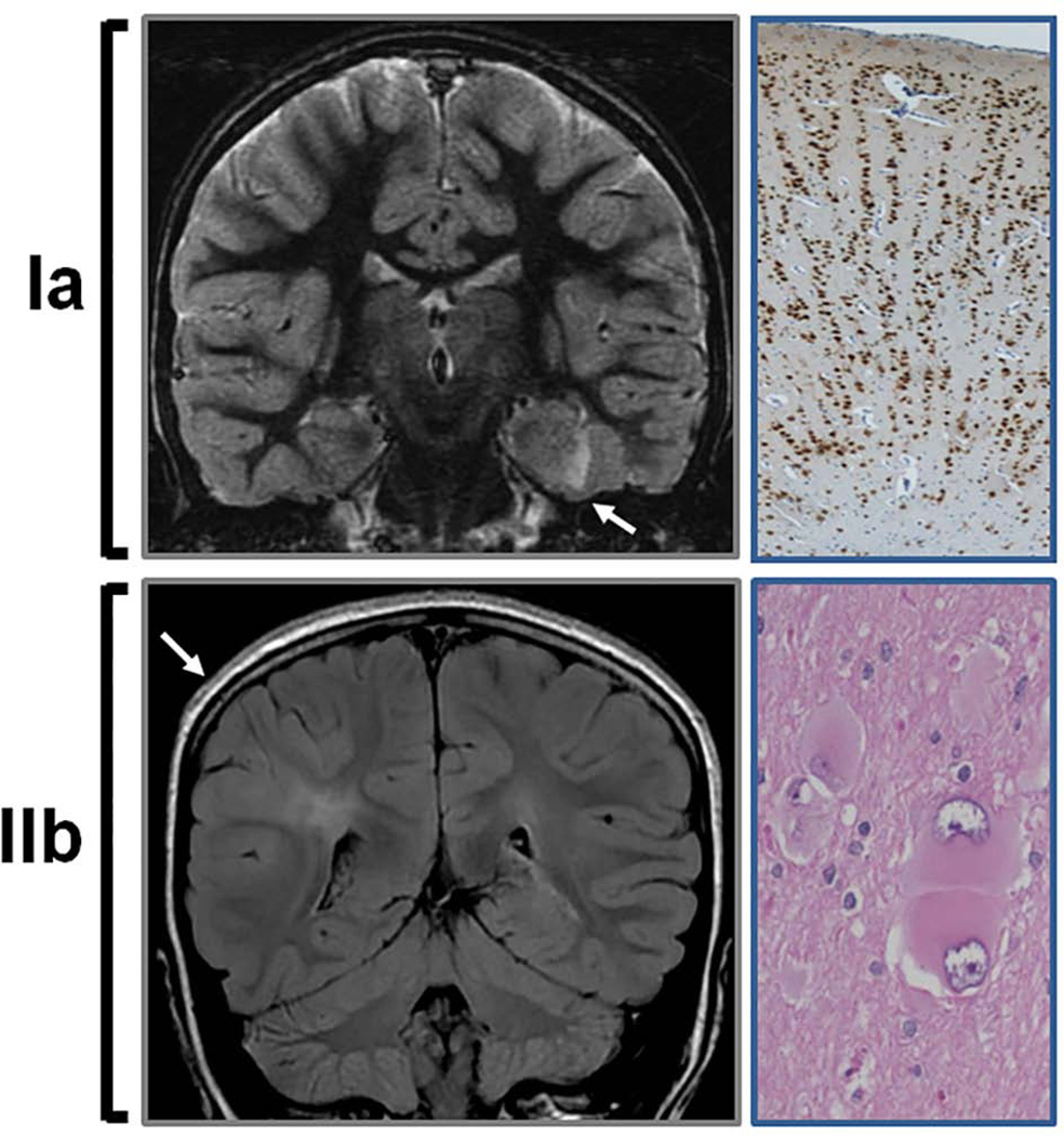
FIGURE 1. Neuroimaging and histological features in FCD type Ia and IIb.
(Upper panel) Coronal T2-weighted MRI illustrating a dysplastic left medial temporal cortex (arrow) corresponding to FCD type Ia. NeuN staining (neuronal marker) of the resected specimen reveals absence of normal lamination and the characteristic radial distribution of neurons (10×). (Lower panel) Coronal fluid-attenuated inversion recovery (FLAIR) MRI illustrates cortical thickening and hyperintense signal in cortex and subcortical regions of the right parietal lobe in FCD type IIb. Note that the subcortical hyperintensity extends to the margin of the right ventricle (transmantle sign). Hematoxylin and eosin staining of the corresponding resected specimen demonstrates the typical balloon cells (20×).

