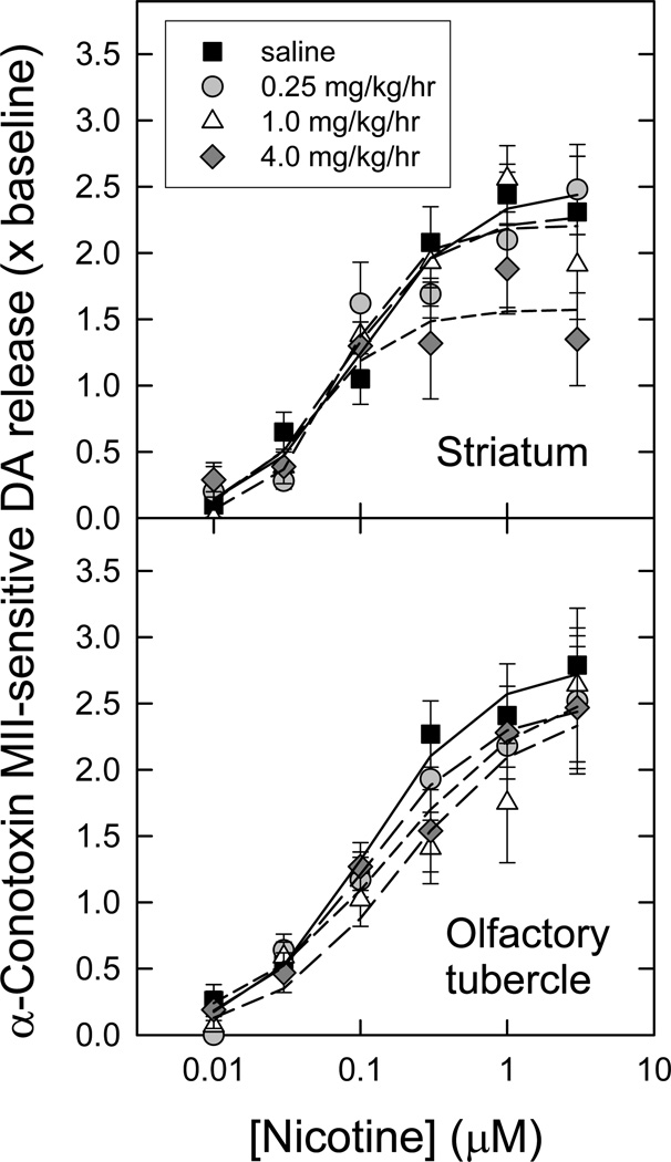
Figure 4. Concentration-response curves for nicotine-evoked [3H]dopamine release mediated by α6β2* nAChR following chronic nicotine administration.
Crude synaptosomes were produced from striatum and olfactory tubercles of animals treated for 10 d with saline (control) or three different nicotine doses as indicated. Synaptosomes were loaded with [3H]-DA, then superfused for 5 min in the presence or absence of 50 nM α-CtxMII just prior to stimulation with a range of nicotine concentrations (10 nM – 3 µM). α-CtxMII-sensitive release (mediated by α6β2* nAChR) of [3H]-DA was calculated by subtracting the resistant fraction from the total (n = 6–11 for each point; points represent mean values ± S.E.M), and is expressed as a multiple of baseline release just before stimulation. The curves for stimulation by nicotine represent the best fits of the data as one saturable component, for each chronic treatment regime, in each region, as described in the Methods. Calculated parameters and statistical analyses are given in Table 2.

