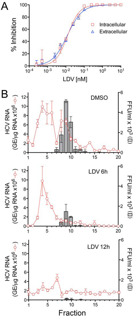
Figure 4.
NS5A inhibitors block intracellular assembly of HCV. (A) Inhibition of intracellular infectious virus (red) and cell-free virus released from infected cells (blue) following 24 hrs treatment with LDV. Intracellular and extracellular virus were quantified in FFU assays. Results are expressed as % inhibition and overlaid for comparison. (B) Rate-zonal centrifugation of cell lysates derived from H77S.3-transfected cells that were either mock-treated (0.0575% DMSO, top panel) or treated with LDV at ~3× the EC90 in the GLuc assay (575pM, bottom panel) for 6 or 12 hrs. Fractions collected from the top of the gradients were tested for HCV RNA by qRT-PCR and for infectivity by FFU assay.

