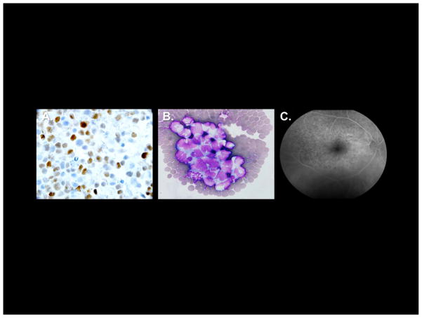
Figure 2. Pathological features of primary central nervous system lymphoma and intraocular lymphoma.
A. High expression of MUM1 by diffuse large B-cell lymphoma cells in a diagnostic specimen of primary central nervous system lymphoma, as demonstrated by immunohistochemistry 1000x). B. Cytological appearance of malignant diffuse large B-cell lymphoma in cerebrospinal fluid from recurrent central nervous system lymphoma. C. Fluorescein angiography showing classic “leopard spots” in Intraocular Lymphoma.

