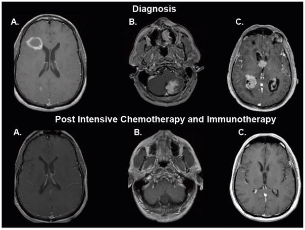
Figure 3. Distinct radiographic presentations of central nrvous system lymphomas and durable responses to intensive chemotherapy and immunotherapy, Without whole brain radiotherapy (T1 axial, post-gadolinium magnetic resonance imaging, at diagnosis and at restaging, at least two months after completion of therapy).
3A. Ring enhancing lesion in the right frontal lobe, adjacent to the lateral ventricle. 3B. Solid enhancing infiltrative mass involving the left cerebellum with evidence of dural attachment. 3C. Diffuse involvement of the ventricular system by nodular, avidly enhancing masses with extension into the surrounding parenchyma.

