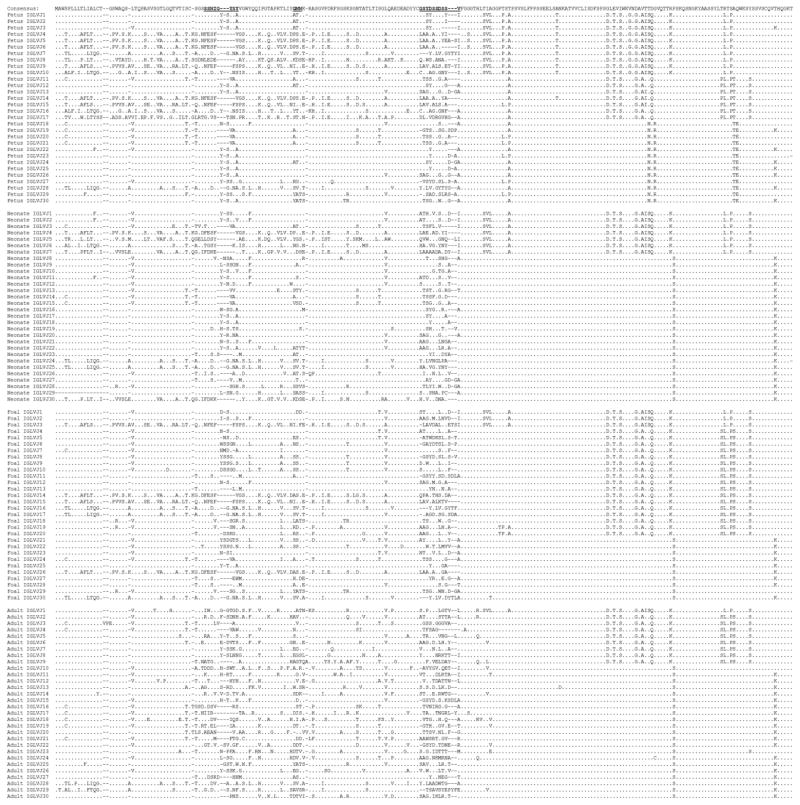
Figure 4. Amino acid alignment of equine immunoglobulin lambda light chain sequences.
Alignment of fetal, neonatal, foal, and adult horse Ig lambda light chain sequences. The 3 complementarity-determining regions are bolded and underlined. Residues identical to the consensus sequence are represented by dots and dashes represent gaps inserted to maximize the alignment.

