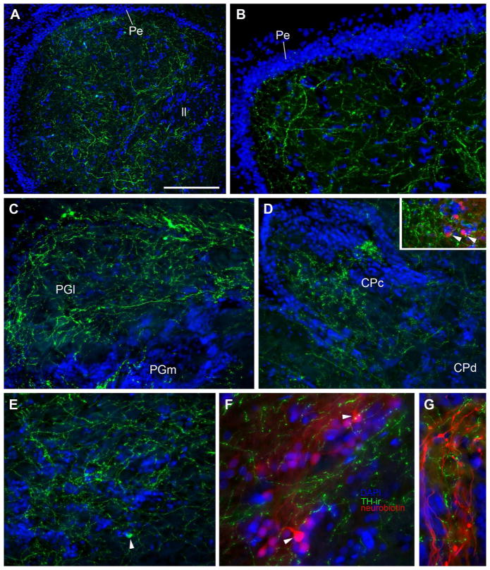
Figure 10.
TH-ir in higher order auditory nuclei; blue is DAPI nuclear stain. A,B: TH-ir fibers and terminals are abundant in the midbrain torus semicircularis (TS). A: Horizontal section through TS. Rostral is to the left, medial is top of the image. B: Transverse section through auditory area centralis of TS. Compact band of nuclei is the periventricular cell layer (Pe) of TS. C: TH-ir projections and varicosities in the lateral (PGl) and medial (PGm) division of nucleus preglomerulosus. Image taken from same section shown in 4C. D: TH-ir terminals in the compact (CPc) and diffuse (CPd) divisions of the central posterior nucleus (auditory thalamus). Inset shows TH-ir terminals on neurobiotin-filled cells (red) in CPc following a bilateral backfill of the saccular branch of VIII. E: A single TH-ir cell (arrowhead) together with dense TH-ir terminals in the hypothalamic anterior tuberal nucleus (AT). Image taken from same section shown in 4C. F: TH-ir terminals on neurobiotin-filled cells (red, arrowheads) in AT following a bilateral backfill of the saccular branch of VIII. AT is also part of the descending vocal motor circuitry and contains reciprocal connections with CP. G: TH-ir terminals are found intermixed with neurobiotin-filled afferents (red) from a saccular backfill in the eminentia granularis. Scale bar in = 200μm in A, 100μm in B–E, 50μm in F and G.

