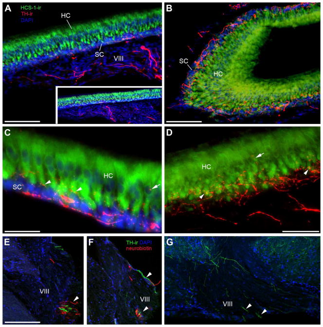
Figure 13.
TH-ir innervation of the saccule, the main endorgan of hearing. A: Section through the saccular epithelium (SE) including the attached branch of the eighth nerve (VIII). The hair cell layer (HC) is delineated using the hair cell specific antibody (HCS-1, green) which labels HC somata and can be distinguished from the basal support cells (SC) labeled by DAPI (blue) alone. Thick and smooth TH-ir fibers (red) course through VIII prior to terminating largely at the base of the HC layer. Inset is lower magnification of same section. B: Horizontal section showing larger varicose TH-ir fibers in the SC and finer terminals in the HC layer of the SE. C,D: High magnification images showing thick TH-ir varicose fibers along the SC layer, fine-caliber terminals (arrowheads) at the base of the HC and less frequently terminals on the central portion of individual hair cells proximal to the nucleus (arrows). E,F: Transverse section through the lateral hindbrain where VIII converges with the CNS. Arrowheads indicate intermingled TH-ir fibers with neurobiotin-labeled fibers from a saccular backfill. G: Horizontal section through the hindbrain; medial is top lateral is bottom of image. Arrowheads show small bundles of TH-ir axons exiting the brain via VIII. Scale bar = 100μm in A and B, 200 μm in set A, 33μm in C, 50μm in D, 200μm in E–G. See supplementary Fig. 4 for magenta-green version of D.

