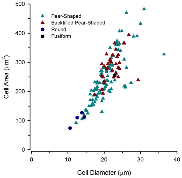
Figure 17.
Size and shape distribution of TH-ir neurons in the periventricular posterior tuberculum that were backfilled after neurobiotin application on the saccular branch of the eighth nerve (red triangles). These cells had an average soma diameter of 22μm and all were classified as pear-shaped. Measurements of other non-backfilled TH-ir neurons within the same sections were taken for comparison.

