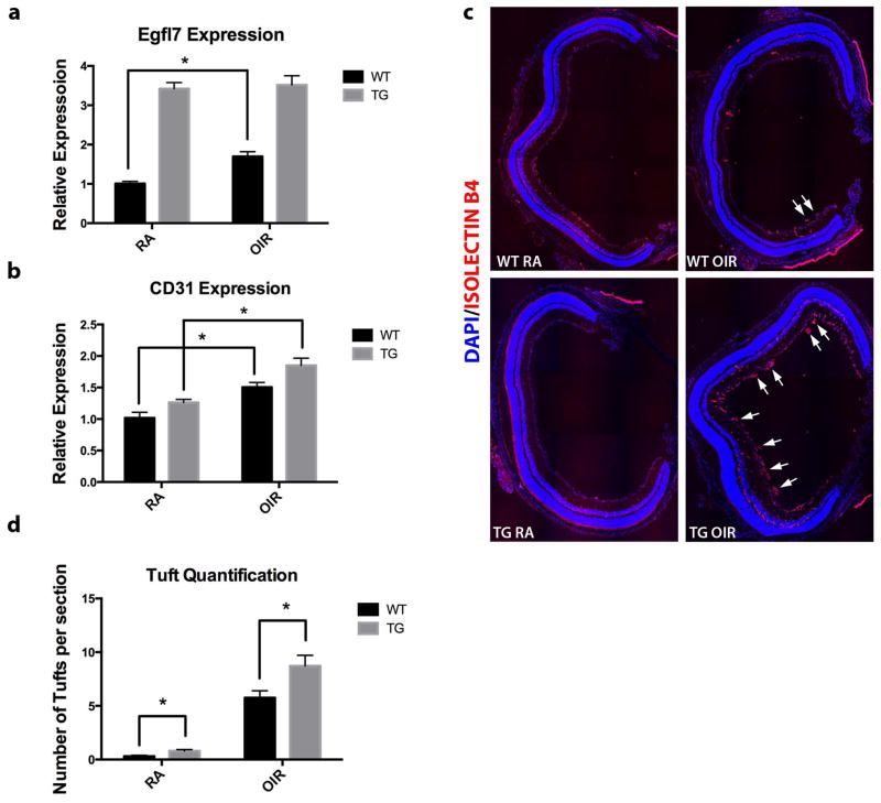
Figure 9. Increased Egfl7 expression contributes to pathological neovascularization in mouse model of ROP.
a) Expression of Egfl7 transcript in WT and Tie2-Egfl7 TG room air and OIR pups at P17. b) Expression of CD31 transcript in WT and Tie2-Egfl7 TG room air and OIR pups at P17. c) Retinas were isolated from P17 pups, sectioned, and stained using DyLight-594-labeled isolectin B4 (red). Representative figures contain composites of 20x tiled images of the entire retina. Arrows indicate pathological vascular tufts. d) Quantification of pathological vascular tufts in P17 retinas (five sections per retina were quantified). Values are represented as mean +/− SEM; *p<0.05. n = 6 for each genotype and each condition.

