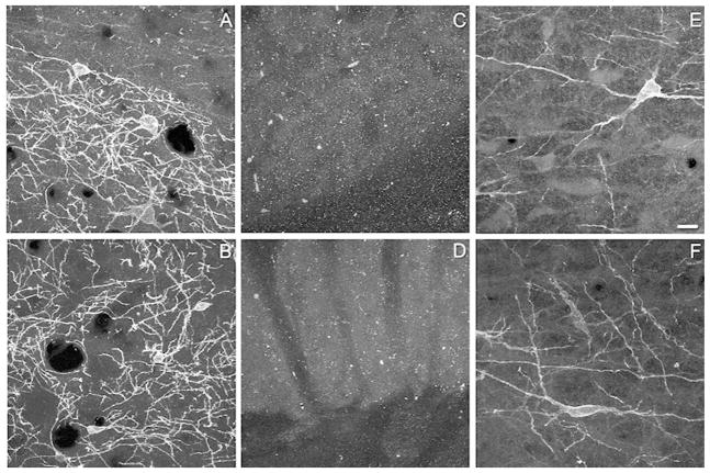
Figure 1.
Representative images demonstrating the specificity of the two neurokinin-1 receptor (NK1R) antibodies. Panels A, C and E were obtained with antisera from Chemicon (Ab5060 lot number LV1378369). Panels B, D and F were obtained using antisera from Sigma-Aldrich (S8305, lot number 067K4885). In the striatum of wild type mice both antibodies produced strong labeling of soma and processes (A,B). Specific labeling was absent in the striatum of a NK1R −/− mouse (C,D). Using the Chemicon antisera, NK1R immunoreactivity was present on the membrane and processes, as well as within the cytoplasm (E) of neurons in the rostral ventromedial medulla (RVM). In contrast, in sections processed with the Sigma antisera (F), NK1R immunoreactivity was largely restricted to the membrane of the soma and processes of neurons in the RVM. Images in panels A–D are projections of four confocal sections taken at an interval of 0.51 or 0.63 microns. Panel E is a projection of three confocal sections taken at an interval of 0.84 microns, while panel F is a projection of four confocal sections taken at an interval of 0.63 microns. Scale bar is 20 microns and applies to each panel.

