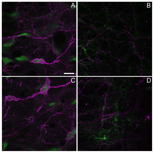
Figure 2.
Neurokinin-1 receptor (NK1R) immunoreactivity is largely neuronal in nature. Distribution of NK1R immunoreactivity (magenta) in the rostral ventromedial medulla of rats four days after an intraplantar injection of (A) saline or (C) complete Freund’s adjuvant (CFA) in the left hind paw. These tissue sections were also processed with an antibody to NeuN (green) to identify neurons. NK1R immunoreactivity rarely co-localized with immunoreactivity for glial fibrillary acidic protein (green) in saline-treated (B) or CFA-treated (D) rats. Images in panels A and C are the projections of a stack of 3–4 confocal sections taken at an interval of 0.76 microns while panels B and D are the projections of 3–4 stacks taken at 0.72 and 0.74 microns respectively. Scale bar for all panels is 20 microns. The gamma was adjusted by 10% for images in panels B and D.

