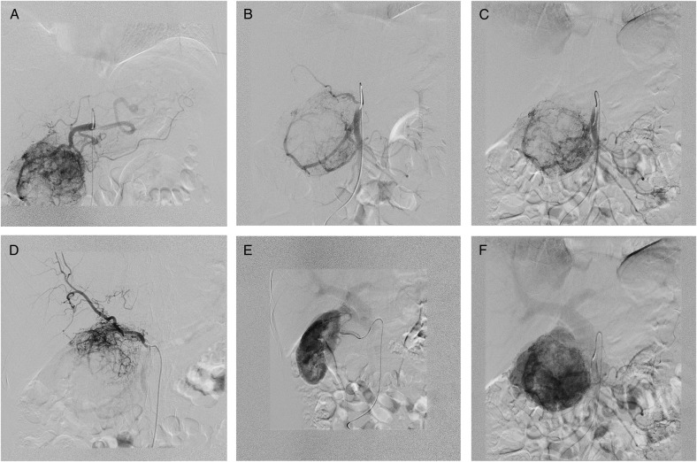
Figure 3:
DSA. (A) GDA supplying the upper portion of the paraganglioma. (B) SMA—tumour blood supply via both IPDA and replaced RHA. (C) Selective IPDA angiography. (D) Selective angiography via replaced RHA. (E) Complementary tumour blood supply via capsular branch of RRA draining into the portal system. (F) PV dilation in the venous phase of angiography.

