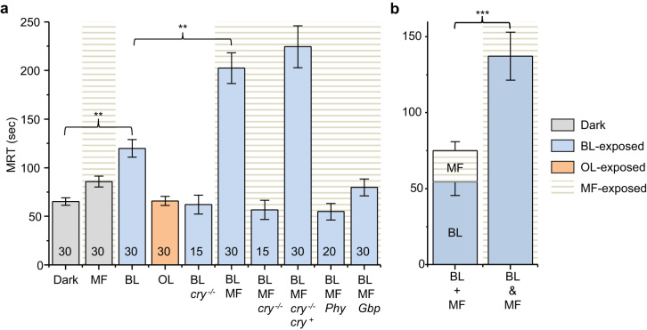
Figure 1. Seizure duration, measured as mean recovery time (MRT), of Drosophila third instar larvae from electric shock.
(a) Third instar larvae developed from embryos exposed to various conditions between 11–19 h after egg laying at 25°C. Colours of bars represent the wavelength of visible light that embryos were exposed to and the presence of a MF is indicated by background horizontal lines. Dark control (Dark); dark + static 100 mT magnetic field (MF); pulsed 470 nm blue light (BL); pulsed 590 nm orange light (OL); pulsed 470 nm + cry03 null (BL/cry−/−); pulsed 470 nm + static 100 mT magnetic field (BL/MF); pulsed 470 nm + static 100 mT + cry03 null (BL/MF/cry−/−); pulsed 470 nm + static 100 mT + cry01/03 null, rescued with expression of DmCRY (BL/MF/cry−/−/cry+); pulsed 470 nm, + static 100 mT + anti-epileptic drug, phenytoin (BL/MF/Phy); pulsed 470 nm, + static 100 mT + anti-epileptic drug, gabapentin (BL/MF/Gbp). All values shown are means ± sem and n is shown in each bar. ** P ≤ 0.01. (b) The combined effect of BL and MF (BL&MF) is significantly larger than the additive effect of BL alone added to MF alone (BL + MF). Values shown are adjusted MRT values, derived by subtracting values obtained in dark controls. *** P ≤ 0.001.

