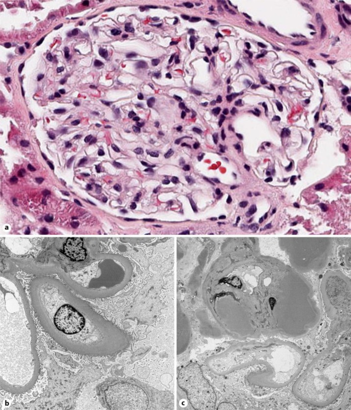
Fig. 1.
a Light microscopy. Photomicrograph of a representative glomerulus showing normal histology (HE. ×40). b Electron microscopy. Diffuse thickening of the lamina densa of the glomerular basement membranes is evident. The epithelial cells are unremarkable, whereas the endothelial swells are swollen with loss of fenestrations (×4,000). c Electron microscopy. Prominent subendothelial hyaline deposits (×2,700).

