
Fig.7.
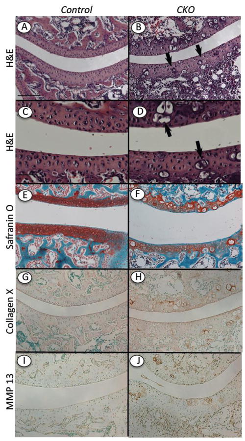
Ectopic hypertrophic-like chondrocytes in mutant articular cartilage. Longitudinal serial sections were prepared of knees from Ext1f/f and Ext1f/f;Col2CreERT mice tamoxifen-injected at P5 and harvested at 2 weeks. (A-F) Staining with H&E (A-D) and Safranin O (E-F) shows that articular cartilage in controls displayed typical cellular organization and strong and uniform matrix staining (A, C, E), whereas articular cartilage in mutants exhibited several hypertrophic-like chondrocytes and uneven matrix staining (B, D, F). (G-J) Immunostaining for collagen X and MMP-13 shows that these hypertrophic markers were essentially undetectable in control articular cartilage (G, I), but were obvious and associated with hypertrophic-like cells in mutant tissue (H, J). Bar= 500 μm (A-B, E-J); 200 μm (C, D).