
Fig. 8.
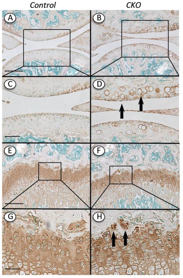
Distribution of Perlecan in articular cartilage and growth plate. Knee sections from Ext1f/f and Ext1f/f;Col2CreERT mice tamoxifen-injected at P5 and harvested at 4 weeks were processed for Perlecan immunostaining using domain 5 antibodies. (A, C, E and G) In controls positive Perlecan staining was evident in top articular cartilage zone and throughout the growth plate. (B, D, F and H) In CKO mutants, positive Perlecan staining was also very strong in the pericellular matrix of the hypertrophic-like chondrocytes in articular cartilage (D, arrows) and the cell clusters present near/within the growth plate reserve zone (H, arrows). Boxed areas in A-B and E-F are shown at higher magnification in C-D and G-H, respectively. Bars= 500 μm (A, B, E, F); 200 μm (C, D, G, H)