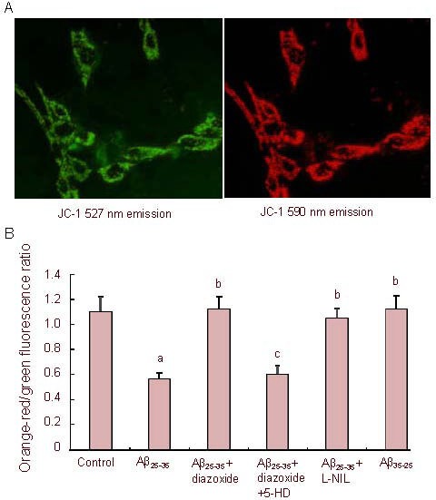
Figure 2.

Effects of amyloid-β peptide (25–35) (Aβ25-35) on mitochondrial membrane potential in PC12 cells.
Cells stained with 5,5’,6,6’-tetrachloro-1,1’,3,3’- tetraethylbenzimidazolcarbocyanine iodide (JC-1) (A, × 400). Green fluorescence (emission maximum at 527 nm) and orange-red fluorescence (emission maximum at 590 nm) are shown.
Cells were exposed to Aβ25-35 (5 µM) or Aβ35-25 (5 µM) in the presence or absence of 1 mM diazoxide (a potassium channel activator) or 1 mM diazoxide plus 500 μM 5-hydroxydecanoate (5-HD, a KATP channel blocker) or 500 μM Nω-nitro-L-arginine (L-NIL, a nitric oxide synthase inhibitor) for 24 hours, followed by staining with JC-1 and laser scan confocal microscopy semi-quantitative analysis of the JC-1 590/527 nm fluorescence ratio (B) (the lower the JC-1 590/527 nm fluorescence ratio is, the higher the mitochondrial membrane potential is).
The data are expressed as mean ± SEM from five independent experiments. aP < 0.01, vs. control; bP < 0.01, vs. Aβ25-35; cP < 0.01, vs. Aβ25-35 + diazoxide (one-way analysis of variance, followed by two-tailed Student's t-test).
