Figure 1.

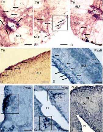
Expression of tyrosine hydroxylase (TH) and transcription factor (Pax6) in the brain of cherry salmon Oncorhynchus masou (immunoperoxidase staining, light microscopy) at different years of age.
(A) Immunolocalization of TH in a spinal cord of a 1-year-old cherry salmon, the arrows show radial glia (RG) cell bodies, localized near a central channel, RG fibers are shown by small arrows on B and C, square marks the sites of fibers with “end feet”, signed by black arrows on B.
(D) TH-immunoreactive RG cell bodies in the superficial layers of the tectum in a 3-month-old cherry salmon (black arrows), and the fibers are shown by small arrows.
(E) Pax6 in tectum of a 6-month-old cherry salmon, the bodies of immunoreactive cells of periventricular layer are shown (solid arrows), the migrated cell bodies (arrows with a cut), the RG fibers (arrow shapes); Pax6- immunoreactive elements in the pretectum on F, thalamus on G and in the cerebellum on H of a 1-year-old cherry salmon, square marks the accumulations of immunoreactive cells, corresponding to pretectal (Pt), dorsal (Dth) and ventral (Vth) thalamic (P1–P3) prosomers; the bodies of Pax6-immunoreactive cells are shown by white arrows, the bodies of migrated cells are shown by black arrows with a cut.
Scale bars: 50 μm for A–D, F; 100 μm for E, G, H. Cc: Central channel, MLF: medial longitudinal fascicle, TeO: optical tectum, Pt: pretectum, Inf: infundibulum; Dth: dorsal thalamus, Vth: ventral thalamus, ML: molecular layer, GL: granular layer, IV: the fourth ventricle.
