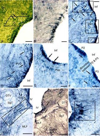
Figure 2.

Expression of tyrosine hydroxylase (TH) in the 3-year-old cherry salmon Oncorhynchus masou brain (immunoperoxidase staining, light microscopy).
(A) Immunoreactive cells and fibers (white arrows) of radial glia (RG) in zona limitans (delineated by parallelepiped). (B) RG under large magnification. (C) Periventricular region of dorsal thalamus (rectangles) delineates the pretectal region (Pt), dorsal thalamic nuclei (Dth) and ventromedial thalamic nuclei (Vmth), the RG fibers are shown by black arrows, the migrating cells are shown by arrows with a cut.
(D) Paraventricular organ (PVO), (E) parvocellular preoptic area (Pop), thin arrows show the RG fibers, black solid arrows indicate periventricular bodies of TH- immunoreactive cells. (F) A border between dorsal forebrain neuromers P2 and P3. (G) Interfascicular segment of the medulla (delineated by rectangle). (H) Ventromedial medullar segment. (I) RG in ventro-medial region of area postrema (delineated by the rectangle).
Scale bars: 100 μm for A, D, G; 20 μm for B; 50 μm for C, E, F, I; 200 μm for H. DL-V: Ventral segment of dorso-lateral region; Zl: zona limitans; Vl: ventro-lateral region; Inf: infundibulum; DTV: descending pathway of N. trigeminus; SgT: secondary gustatory tract; IX–X: nuclei of glossopharyngeal nerve and vagus; MLF: medial longitudinal fascicle; MRF: medial reticular formation; IV: the fourth ventricle; AP: area postrema.
