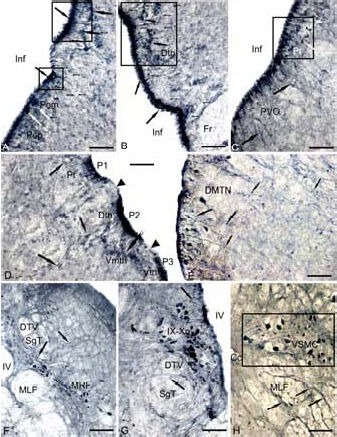
Figure 3.

Expression of gamma amino butyric acid (GABA) in the 3-year-old cherry salmon Oncorhynchus masou brain (immunoperoxidase staining, light microscopy).
(A) Clusters of immunoreactive radial glia (RG) (delineated by squares) in preoptical region, the neurons of a magnocellular nucleus (Pom) are shown by white arrows, the periventricular cells bodies by black arrows, RG fibers by a figure like arrows, and the migrated cells by arrows with a cut.
(B) RG in a region of dorsal thalamus. (C) Paraventricular organ, the cells of anterior tuberal nucleus (delineated by a square) are indicated by white arrows. (D) RG in the content of forebrain prosomers P1, P2 and P3 without GABA immunolabeling on the border (black edges of black arrows). RG fibers (black arrows) and migrated cells in the content of pretectum (Pt), dorsal thalamus (Dth) and ventro-medial nuclei of thalamus (Vmth) are shown by arrows with a cut, (E) RG in the content of dorsomedial tegmentum by black arrows, and white arrows for large neurons of DMTN. (F) RG in the interfascicular area (black arrows) and immunoreactive cells MRF (white arrows). (G) RG on the territory of nuclei of IX–X craniocerebral nerves. (H) RG on the territory of the spinal cord (neuronal bodies are shown by white arrows).
Scale bars: 100 μm for A–D, G, H; 200 μm for E, F. Fr: Fasciclus retroflexus; Inf: infundibulum; PVO: paraventricular organ; DMTN: dorsomedial tegmental nuclei; Vth: ventral thalamu; DTV: descending pathway of N. trigeminus, SgT: secondary gustatory tract, MLF: medial longitudinal fascicle; MRF: medial reticular formation; IV: the fourth ventricle; IX–Xn: nuclei of glossopharyngeal nerve and vagus; Cc: central channel; VSMC: ventral spinal motor column.
