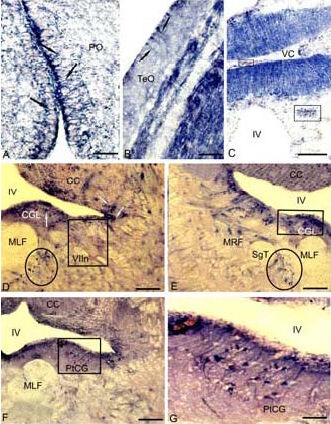
Figure 4.

Localization of nicotinamide adenine dinucleotide phosphate-diaphorase (NADPH-d) in the 3-year-old cherry salmon Oncorhynchus masou brain (histochemical staining; light microscopy).
(A) Radial glia (RG) in a parvocellular preoptical region (black arrows), in superficial layers of tectum on B, in medial part of valvula cerebelli (VC) on C, complexes of vessels and perivascular glia are delineated by the rectangle.
(D) RG (white arrows) in a central grey layer (CGL) of subventricular region in the territory of nucleus of facial nerve (VIIn) (NADPH-d-negative nucleus VIIn is delineated by a rectangle), accumulation of RG adjacent to medial longitudinal fascicle (in an oval).
(E) RG clusters CGL in the territory of reticular formation, a cluster of NADPH-d-positive cells of RG (in a square). (F) RG in the territory of posttrigeminal group of a central grey layer (in a square). (G) Fragment inside a square in F on a large magnification.
Scale bars: 50 μm for A, G; 20 μm for B; 200 μm for C–F. PO: Preoptical region; CC: corpus cerebellum; MLF: medial longitudinal fascicle; MRF: medial reticular formation; IV: the fourth ventricle; SgT: secondary gustatory tract; PtCG: posttrigeminal group of central grey layer; TeO: tectum opticum.
