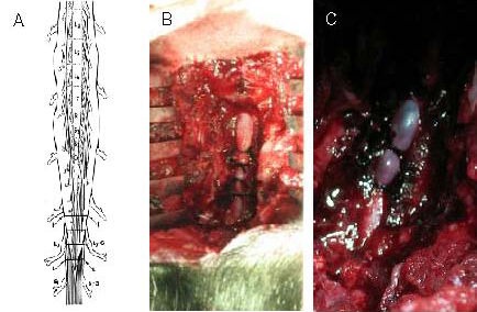
Figure 3.

Procedure for the establishment of multiple protracted cauda equina constrictions model.
(A) Schematic drawing depicting the position of four tightened constrictions (about 2.0 mm wide) around the cauda equina (L, L). L7–G and S1–G point to the corresponding dorsal root ganglia to produce acute and severe compression, and the entire cauda equina was constricted by 50–75% by the first tightened constriction. The lower cauda equina was constricted by 25–50% by the other three tightened constrictions.
(B) Protracted multiple cauda equina constrictions during surgery on an experimental animal.
(C) Severe arterial narrowing at the level of the constriction and venous congestion of the nerve roots and dura mater of the corresponding lumbar and sacral levels were present after 48 hours of multiple cauda equina constrictions.
