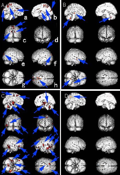
Figure 1.

Activation/deactivation differences in functional areas of the brain between stroke patients and healthy controls following acupuncture at Waiguan (TE5).
Red region (arrows): differences in activation/deactivation.
a: Left sagittal plane of the median brain; b: right sagittal plane of the median brain; c: posterior plane of the brain; d: anterior plane of the brain; e: left view of the brain; f: right view of the brain; g: normal inferior of the brain; h: apical view of the brain (a–h in all pictures represent the same plane as Figure A).
(A) Enhanced activated brain areas in ischemic stroke patients compared with normal controls.
(B) Attenuated activated brain areas in ischemic stroke patients compared with normal controls.
(C) Enhanced deactivated brain areas in ischemic stroke patients compared with normal controls.
(D) Attenuated deactivated brain areas in ischemic stroke patients compared with normal controls.
