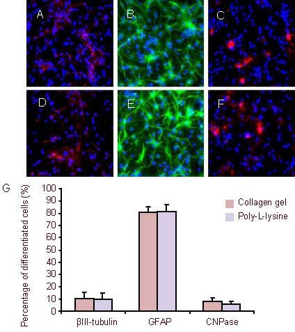
Figure 7.

After culturing for 7 days, the neural stem cells were induced to differentiate (immunofluorescence staining, × 400).
Fluorescent photomicrographs represent differentiated cell phenotypes from neural stem cells cultured in three-dimensional collagen gels (A–C) and in suspension (D–F): βIII-tubulin-positive neurons (A and D), GFAP (B and E, green) and CNPase (C and F, red). Nuclei were stained with 4’,6-diamidino-2-phenylindole (blue).
(G) Percentage of differentiated cells. Data are expressed as mean ± SD of three replicates (one-way analysis of variance, two-sample t-test).
CNPase: 2’,3’-Cyclic-nucleotide 3’-phosphodiesterase; GFAP: glial fibrillary acidic protein.
