Figure 1.
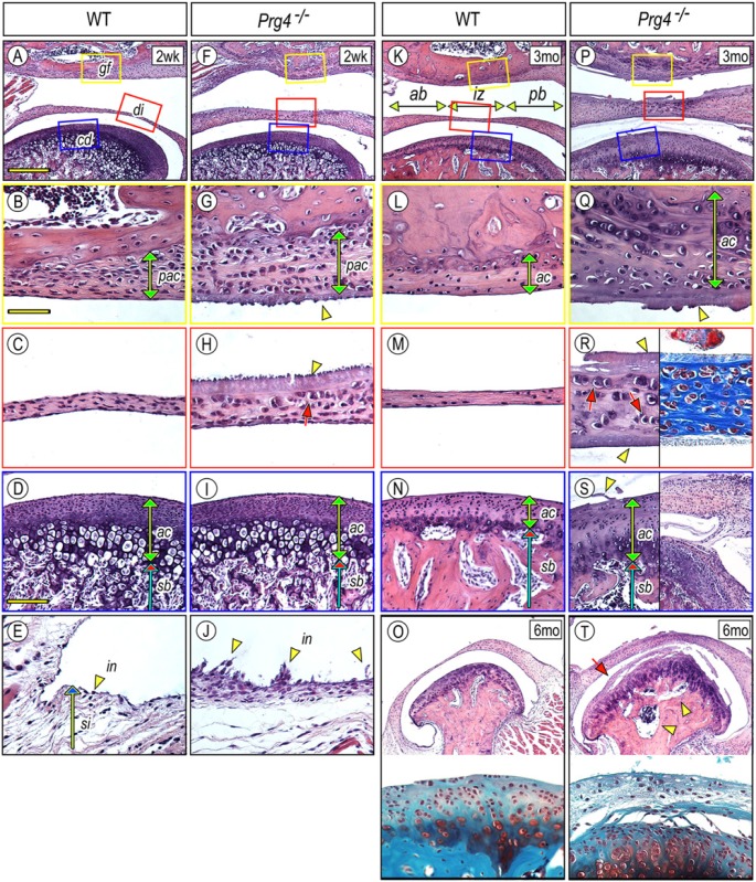
Glenoid fossa, articular disc, synovial membrane, and mandibular condyle are defective in Prg4–/– TMJs. TMJs from 2-week (A-J), 3-month (K-N, P-S), and 6-month-old (O, T) control (A-E, K-O) and Prg4–/– (F-J, P-T) mice were analyzed by histology. Hematoxylin and eosin (H&E) (A-T), Safranin O/Fast green (O, T, lower panel), and Masson’s trichrome (R, right panel) staining. Magnified sagittal views of glenoid fossa (B, G, L, Q), articular disc (C, H, M, R), and mandibular condyle (D, I, N, S, O, T) were obtained from each colored boxed area in the top panel of each line or corresponding sections. Note that H&E- or Masson’s trichrome-stained substance covers the surface of the glenoid fossa, disc, and condyle (G, H, Q, R, S arrowhead). There are no clear signs of inflammatory cell invasion into the synovial membrane (E, J). Scale bars: 250 µm in A for A, F, K, P, O, T; 65 µm in B for B, C, E, G, H, J, L, M, Q, R; and 150 µm in D for D, I, N, S. gf, glenoid fossa; di, disc; cd, condyle; sb, subchondral bone; pac, presumptive articular cartilage; ac, articular cartilage; in, intima; si, subintima; ab, anterior band; iz, intermediate zone; pb, posterior band.

