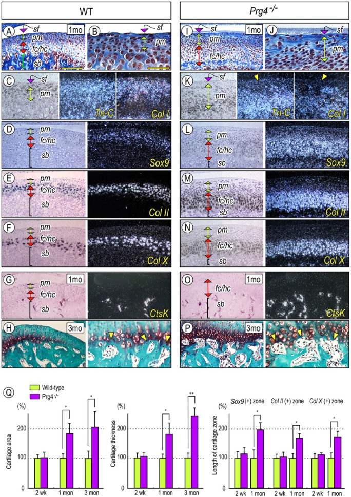
Figure 2.
Prg4 mutants display defective superficial layer, abnormal subchondral bone, and increased chondrogenesis in the mandibular condyle. Serial parasagittal sections from 1-month (A-G, I-O) and 3-month-old (H, P) control (A-H) and Prg4–/– (I-P) TMJs. Sections were processed for Masson’s trichrome (A, B, I, J) and Safranin O/Fast green (H, P) staining and in situ hybridization (C-G, K-O) with isotope-labeled riboprobes for tenascin-C (Tn-C) (C, K), type I collagen (Col I) (C, K), Sox 9 (D, L), type II collagen (Col II) (E, M), type X collagen (Col X) (F, N), and Catepsin K (CtsK) (G, O). (Q) For quantification of the Safranin O-staining area and the thickness of chondrocyte zones, Sox9-expressing, Col II-expressing, and Col X-expressing zones along the longitudinal axis were measured from randomly selected 3-5 sections per sample (n = 3 for each mouse/each group) and presented as average ± SD. Control data were set as 100%. p values less than .05 were considered as statistically significant (*p < .01, **p < .005). Scale bars: 250 µm in A for A, D-H, I, L-P; 55 µm in B for B, C, H (right panel), J, K, and P (right panel). sf, superficial layer; pm, polymorphic layer; fc, flattened chondrocyte zone; hc, hypertrophic chondrocyte zone; sb, subchondral bone.

