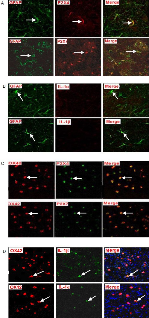
Figure 6.

Double fluorescence staining for the P2X4 receptor, P2X7 receptor, interleukin (IL)-1α and IL-1β expression in astrocytes and microglia in hippocampal slices subjected to oxygen-glucose deprivation (confocal laser scanning microscope, × 100).
At 75 minutes after oxygen-glucose deprivation, in green astrocytes (Cy2-stained; glial fibrillary acidic protein, GFAP), P2X4 and P2X7 receptors were stained red with Cy3. After merging the two images, yellow staining appeared (arrows; A) indicating colocalization. IL-1α and IL-1β were not stained with Cy3. After merging of these images, no colocalization was observed (arrows; B). In red microglia (Cy3 stained; OX42), P2X4 receptor, P2X7 receptor, IL-1α and IL-1β all stained green with Cy2, and after merging of the images yellow double staining appeared (arrows; C, D), indicating colocalization.
