


Figure 4.
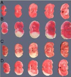
Representative photographs of coronal brain sections stained with TTC.
The infarct lesions are shown as white areas.
(A) TTC staining of coronal sections in normal rat brain, no infarct lesions are visible.
(B) TTC staining of coronal sections in MCAO rats (control group) without cell administration, infarct lesions are obvious.
(C) TTC staining of coronal sections in MCAO rats with BMCs administration, infarct lesions are significantly decreased compared with the control group.
(D) TTC staining of coronal sections in MCAO rats with BMRCs administration, infarct lesions are significantly decreased compared with the BMCs group.
TTC: 2,3,5-Triphenyltetra zolium chloride; BMCs: bone marrow cells; BMRCs: bone marrow regenerative cells; MCAO: middle cerebral artery occlusion.