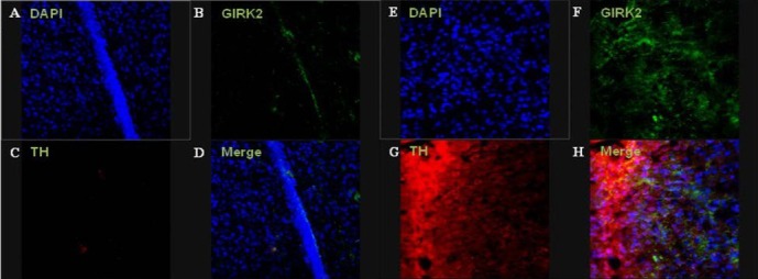
Figure 9.

Immunofluorescence staining of the lesioned striatum at 12 weeks after cell transplantation.
Images showing colocalization of tyrosine hydroxylase (TH)- and G protein-activated inward rectifier potassium channel 2 (GIRK2)-positive cells were partially observed in the injection site of the human brain-derived neural stem cell (HB-NSC) group. Immunohistochemical results of GIRK2 (green; B, F), TH (red; C, G), and merged images (D, H). The control (A–D) and human brain-derived neural stem cell groups (E–H) are shown (all images: × 400 magnification).
