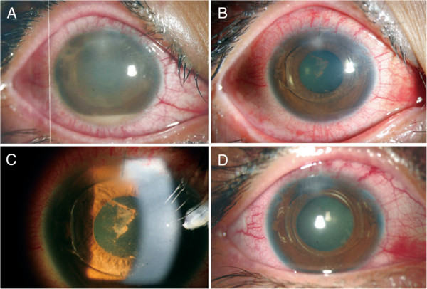
Figure 1.

Anterior segment photographs. A: Right eye shows conjunctival injection, corneal edema with infiltrates at the corneal incision site, membrane formation around the phakic intraocular lens (pIOL), and a 1.5 mm hypopyon 2 days after implantation. B, C: The day after anterior chamber irrigation with intracameral antibiotic injection, the eye shows marked decreased inflammation and corneal edema. D: At 2 weeks after intravitreal antibiotics injection, uncorrected distant visual acuity improved to 20/40 with a well-centered pIOL.
