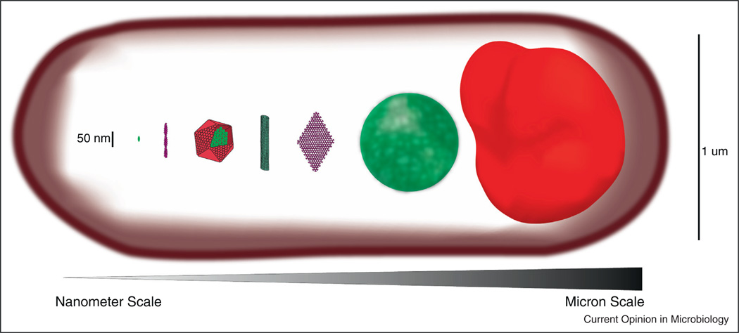
Figure 1.
Various mesoscale self-assembling structures drawn to scale in comparison to a single monomer. From left to right, typical scale of a monomeric scaffold like the yeast protein Ste5, an actin filament, a carboxysome shell housing the enzymes of carbon fixation, a microtubule, a chemotaxis array, a low complexity sequence hydrogel, a purinosome. All structures are assembled within a typical E. coli cell (also drawn to scale).

