
Figure 5.
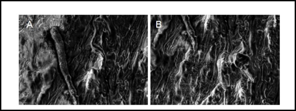
Scanning electron microscopy images showing the ultrastructure of normal human sciatic nerve before and after stress relaxation and creep experiments (× 2 000).
Normal sciatic nerve (A): Fibers were arranged in rows, axons and other components were clearly seen. Sciatic nerve after stress relaxation and creep experiments (B): Fibers were arranged in rows, axons and other components were still clearly visible, with no obvious change.