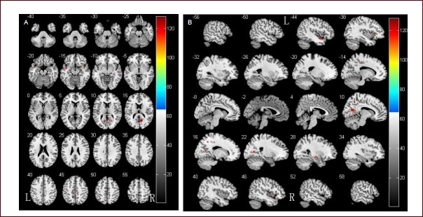
Figure 1.

Comparison of gray matter volume in Parkinson's disease patients with and without dementia.
(A) Results of statistical analysis are represented as pseudo-color on axial template brain mapping in the Montreal Neurological Institute (MNI) standard coordinate.
(B) Results of statistical analysis are represented as pseudo-color on sagittal template brain mapping in the MNI standard coordinate.
Two-sample t-test showed that brain gray matter volume was significantly reduced in Parkinson's disease dementia patients.
R: Right; L: left.
