Figure 1.

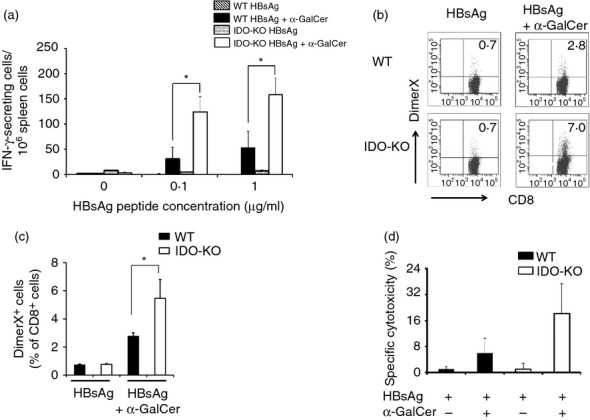
The induction of an hepatitis B virus surface antigen (HBsAg) S28–39-specific T cytotoxic type 1 response and CD8+ T cells in wild-type (WT) and indoleamine 2,3-dioxygenase knockout (IDO-KO) mice immunized with HBsAg alone or in combination with α-galactosylceramide (α-GalCer). Splenocytes were isolated from the animals 7 days after the immunization. (a) These cells were stimulated ex vivo with the HBsAg S28–39 peptide and monitored for interferon-γ (IFN-γ)-secreting cells by means of an ELISPOT assay. Results are shown as mean ± SEM (four or five mice/group) for three independent experiments. (b) Induction of HBsAg-specific CD8+ T cells was assessed by flow cytometric analysis using fluorescent dimeric H-2Ld-HBsAg S28–39 complexes (DimerX). FACS profiles are shown for WT and IDO-KO mice immunized with either HBsAg or HBsAg plus α-GalCer. (c) Quantitative data on the frequency of dimeric H-2Ld-HBsAg-positive cell populations (mean ± SEM, n = 4). (d) Isolated effector cells (CD8+ T cells) from WT and IDO-KO mice immunized with HBsAg and α-GalCer were incubated for 4 hr with CFSE-labelled target cells (preS1-transfected P815 cells) at an effector to target cell ratio of 20 : 1. The percentage of specific cytotoxicity was calculated by subtracting the percentage of P815 cells (HBsAg-negative) effector cell cytotoxicity from that for the preS1-transfected P815 cells (HBsAg-positive). Spontaneous release was always < 20% of the total. Each data point and error bar represents the mean and SEM, respectively, of results for triplicate samples. *Statistically significant differences.
