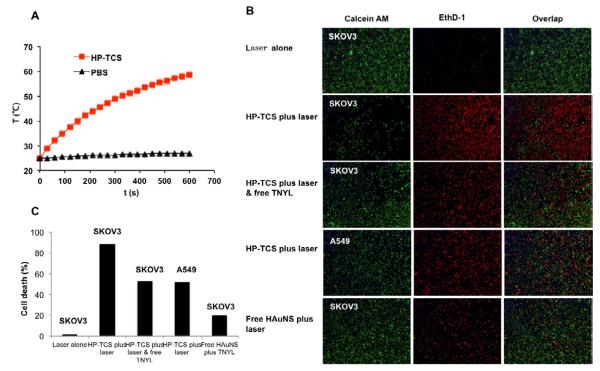
Figure 4.
(A) The temperature changes in aqueous solutions containing HP-TCS micelles (0.5 mg/mL) after exposure to NIR light at an output power of 1.5W. PBS=phosphate buffered saline. (B) Photothermal ablation in SKOV3 and A549 cells with HP-TCS micelle, HAuNS alone or HP-TCS micelles plus free TNYL (blocking). NIR laser was delivered at an output power of 1.5 W for 3 min. Cells were stained with calcein AM (green) and EthD-1 (red) for visualization of live and dead cells, respectively. (C) The quantitative analysis based on the imaging in (B) by the software “Image J”.

