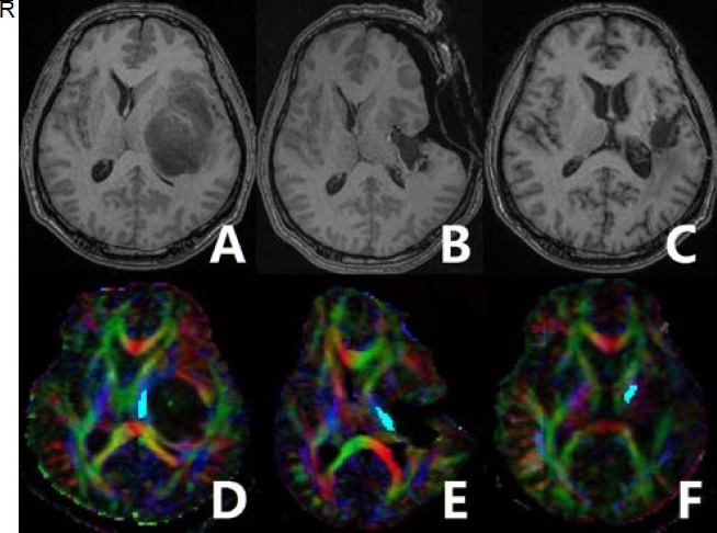
Figure 2.

A 39-year-old male olibgodendroglioma patient with long-term motor function deficits.
Preoperative T1-weighted imaging (T1WI) (A) showing a lesion located in the left insular lobe. The left internal capsule was compressed. Intraoperative (B) and 12-month follow-up (C) T1WI showing that the tumor was resected. Preoperative diffusion tensor imaging-based fiber tracking showing that the corticospinal tract was markedly displaced to the contralateral side (D). Fiber tracking based on preoperative (D), intraoperative (E), and follow-up (F) diffusion tensor imaging was performed.
Red represents a predominant left-right anisotropic diffusion gradient; green represents an anterior-posterior gradient; and blue represents a superior-inferior gradient orientation. R: Right.
