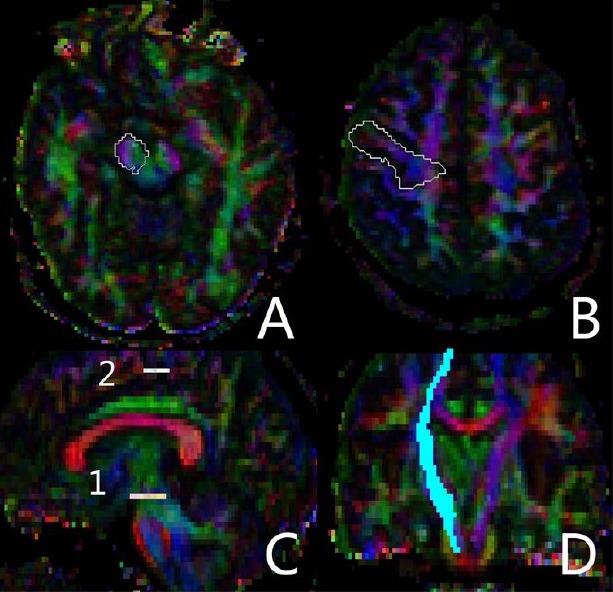
Figure 3.

Identification of the region of interest in diffusion tensor images during fractional anisotropy measurement.
In the two regions of interest, one seed region included the ipsilateral cerebral peduncle in an axial plane at the level of the decussation of the superior cerebellar peduncle (A), and the second seed region was placed on the precentral gyrus (B); Sagittal view showing the level of the two regions of interest (C), and coronal view showing the reconstructed corticospinal tract (D).
