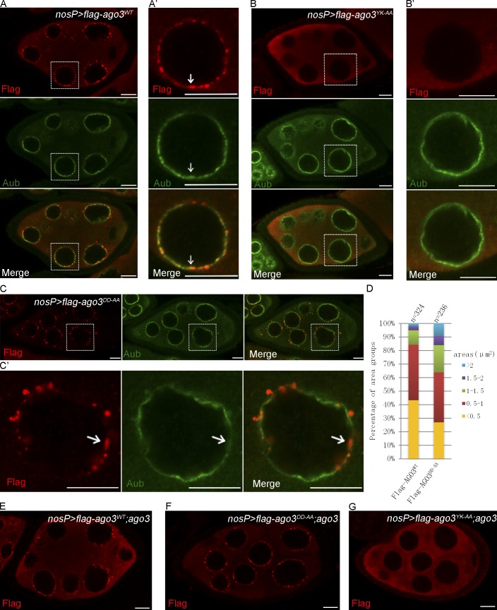
Figure 4.
The Slicer activity of AGO3 is dispensable for its subcellular localization. (A–C′) Ovaries expressing Flag-AGO3WT (A and A′), Flag-AGO3YK-AA (B and B′), and Flag-AGO3DD-AA (C and C′) driven by nosP-gal4:vp16 were stained with anti-Flag (red) and anti-Aub (green) antibodies. A′, B′, and C′ show enlarged images of the boxed portions in A, B, and C, respectively. The arrows indicate the nuage where AGO3 is expressed (A′ and C′) or not (B′). (D) The quantification of the aggregate areas of Flag-AGO3WT and Flag-AGO3DD-AA. The area values were divided into five groups: <0.5, 0.5–1.0, 1.0–1.5, 1.5–2.0, and >2.0 µm2. (E–G) Ovaries from uasp-flag-ago3WT; nosP-gal4:vp16, ago3 (E), uasp-flag-ago3DD-AA; nosP-gal4:vp16, ago3 (F), and uasp-flag-ago3YK-AA; nosP-gal4:vp16, ago3 (G) were stained with anti-Flag (red) antibody. Bars, 10 µm.

