Figure 1.

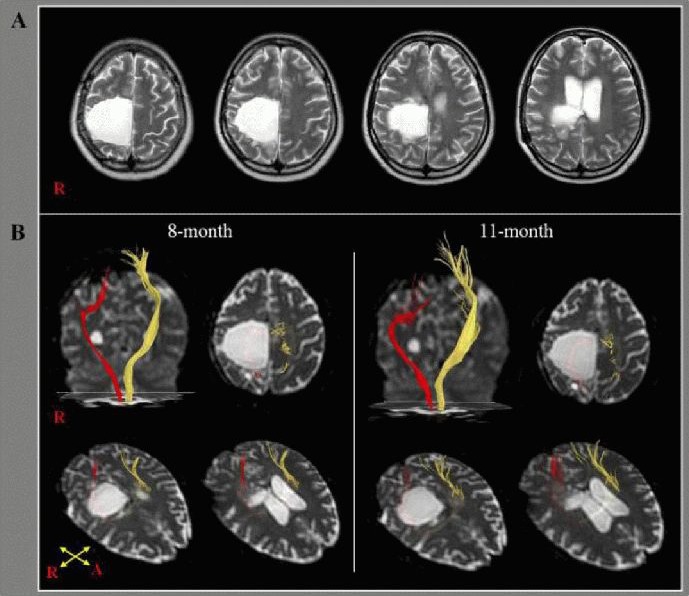
T2-weighted brain MR and diffusion tensor tractography in a 32-year-old, right-handed female with right intracerebral hemorrhage.
(A) T2-weighted brain MR images 8 months after onset showing a large leukomalactic lesion in the right frontoparietal cortex including the primary motor cortex, centrum semiovale, and corona radiate. R: Right.
(B) Diffusion tensor tractography of the corticospinal tract (CST). The CST of the left hemisphere (yellow) originated from the primary sensorimotor cortex and descended through the known CST pathway at 8 and 11 months after stroke. By contrast, the right CST (red) originated from the posterior parietal cortex and descended through the posterior margin of the leukomalactic lesion at 8 months. No significant change was observed at 11 months. R: Right; A: anterior.
