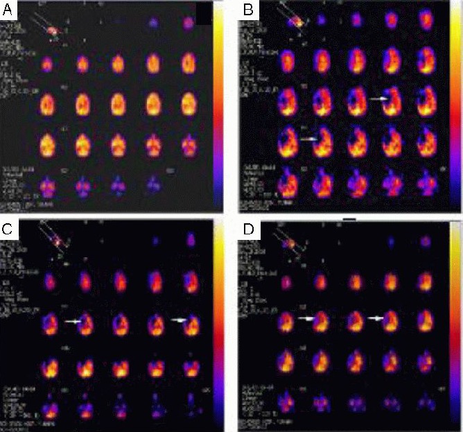
Figure 4.

Changes of cerebral blood flow in rhesus macaques with brain ischemia (SPECT of one rhesus macaque as representative).
(A) 1 day prior to surgery; (B) 3 days post-surgery; (C) 10 days post-surgery; (D) 60 days post-surgery. Arrows represent the blood supply of the M1 segment of the right middle cerebral artery. The blue region reflects that the radioactivity of regional brain tissues was significantly reduced or even lost, indicating ischemic focus. The red region indicates normal radioactivity of regional brain tissues, suggesting normal blood flow perfusion. The yellow region (even white) represents higher radioactivity of regional brain tissues than normal brain tissues, indicating increased blood flow. At 3 days post-surgery, ischemia was evident in the injury area. Up to 60 days, the ischemic region was significantly diminished, but not restored to normal.
