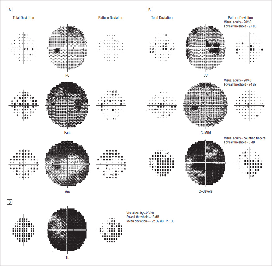
Figure 1.
Classifications for optic nerve visual field abnormalities. A, Nerve fiber bundle abnormalities: paracentral (PC), partial arcuate (Parc), and arcuate (Arc). B, Central abnormalities: centrocecal (CC), central (C)—Mild, and C —Severe. C, Severe abnormalities: total loss (TL) of vision. The total deviation plot is on the left and the pattern deviation plot is on the right of each gray scale for all visual fields.

