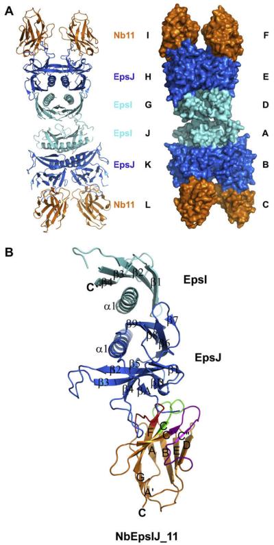
Fig. 2.
The structure of V. vulnificus EpsI:EpsJ in complex with Nb11. (A) View of the unit cell with four EpsI:EpsJ:Nb11 ternary complexes. EpsI, light blue; EpsJ, blue; and Nb11, orange. The cartoon representation of the unit cell is seen on the left, and the surface representation is seen on the right. The chain names in the structure are next to their respective components of the unit cell. (B) General architecture and secondary structure elements of the EpsI:EpsJ:Nb11 ternary complex. EpsI, light blue; EpsJ, blue; and Nb11, orange. Nb11 is further colored by CDR, with CDR1 being green, CDR2 purple, and CDR3 red. Each α-helix and β-strand has been labeled according to its order in the protein. (For interpretation of color mentioned in this figure the reader is referred to the web version of the article.)

