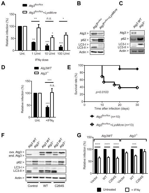
Figure 4. Atg3 is required for the control of T. gondii infection.
(A) Flow cytometry analysis for T. gondii infection at 24 hpi (MOI=1) in Atg3flox/flox+/−LysMcre BMDMs +/− 24 hr pre-treatment of IFNγ at the indicated doses. (B) Protein blot for untreated or uninfected Atg3flox/flox+/−LysMcre BMDMs. (C) Protein blot for WT and Atg3−/− MEF. (D) Same analysis as shown in (A) for the WT and Atg3−/− MEF. (E) Survival curves of Atg3flox/flox+/−LysMcre mice after intraperitoneal inoculation with T. gondii. (F) Protein blot for untreated or uninfected samples as shown in (G). (G) Same analysis as shown in (A) for WT and Atg3−/− MEFs transduced with control, WT Atg3, or its enzyme-null mutant (C264S) +/− 24 hr pre-treatment of 100 U/ml of IFNγ. Statistical analysis by 1-ANOVA with Tukey post test or Log-rank (Mantel-Cox) test. n.s.: not significant (p>0.05), **: p<0.01, ***: p<0.001, ****: p<0.0001. Combined data as average ± SEM. See also Figure S3.

