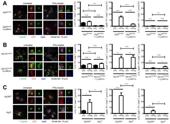
Figure 6. Atg5 and Atg3, but not Atg14L, are required for the localization of LC3 and Irga6 on the PVM of T. gondii.
Representative images (left) and quantitation (right) of immunofluorescence for T. gondii, LC3, and Irga6 in (A) Atg5flox/flox+/−LysMcre and (B) Atg14Lflox/flox+/−LysMcre BMDMs and (C) Atg3 WT and Atg3−/− MEFs at 2 hpi (MOI=1) of T. gondii infection +/− 24 hr pre-treatment of 100 U/ml of IFNγ. At least 100 cells infected T. gondii were analyzed for quantitation. Statistical analysis by 1-ANOVA with Tukey post test. n.s.: not significant (p>0.05), **: p<0.01, ***: p<0.001, ****: p<0.0001. Combined data as average ± SEM.

