. 2014 Jul 29;3:e03025. doi: 10.7554/eLife.03025

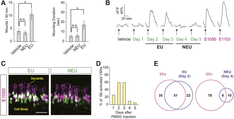
Figure 1. Sulfated estrogens mimic the activity of estrus signal in urine.

(A) The number (left) and duration (right) of mounting behavior of sexually naïve males toward ovariectomized females painted with vehicle (n = 16), non-estrus urine (NEU; n = 14) or estrus urine (EU; n = 22). EU and NEU were collected 2 and 4 days, respectively, after PMSG injection to induce estrus. Error bars, SEM; *p<0.05 (Mann–Whitney test). (B) Traces showing GCaMP2 responses of a representative cell to urine samples collected from females 1 to 5 days after PMSG injection and SEs. Arrows indicate the onset of stimulus delivery. (C) Representative images of the VNO slice response pattern to E1050 (magenta) and urine (green). Scale bar, 50 μm. (D) Bar graph showing the percentage of SE-activated VSNs (n = 86; 3 slices) that are also activated by female urine samples. (E) Venn diagrams showing the overlap between VSNs responding to SEs and those activated by EU or NEU.

DOI: http://dx.doi.org/10.7554/eLife.03025.003

Figure 1.

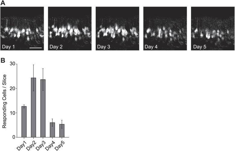
Figure 1—figure supplement 1. Activation of the VSNs by female mouse urine.

Figure 1—figure supplement 1.

(A) Representative images of the VNO slice response pattern to urine samples collected from females 1 to 5 days after PMSG injection. Scale bar, 50 μm. (B) Bar graph showing the number of responding cells to urine samples collected from females 1 to 5 days after PMSG injection in each slice (3 slices). Error bars, SEM.
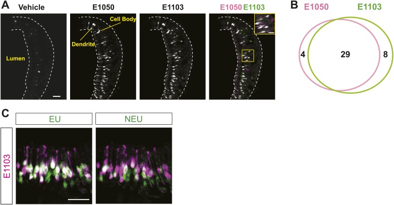
Figure 1—figure supplement 2. Activation of the VSNs by sulfated estrogen E1050 and E1103.

Figure 1—figure supplement 2.

(A) Representative images of a GCaMP2 VNO slice responding to vehicle, 100 nM E1050 and E1103. Bright cells are the activated neurons. VSNs responding to E1050 (magenta) or E1103 (green) are color-coded in the right panel to show overlap (white). Scale bar, 50 μm. (B) Venn diagram of the overlap of VSNs responding to 100 nM E1050 and E1103. (C) Representative images of the VNO slice response pattern to E1103 and urine samples collected from females 2 or 4 days after PMSG injection. VSNs responding to E1103 (magenta) or urine samples (green) are color-coded to show overlap (white). Scale bar, 50 μm.