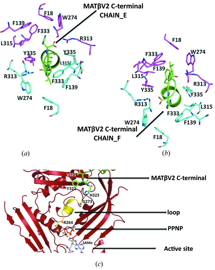
Figure 4.
Close view of the tunnel created at the interface of the MATα2 dimer. (a) Stick representation of the MATα2 residues at the dimer interface, side chains involved in the interaction with MATβV2 (chain_E in green) are coloured in cyan, the symmetry residues are shown in magenta. (b) Side chains involved in the interaction with MATβV2 (chain_F in green) are coloured in magenta, the symmetry residues are shown in cyan. Note that between the conformation represented in (a) and (b) there is a twofold symmetry. (c) Cartoon representation of the MATα2 monomer; the loop that connects the active site with the buried tail of MATβV2 is highlighted in yellow.

