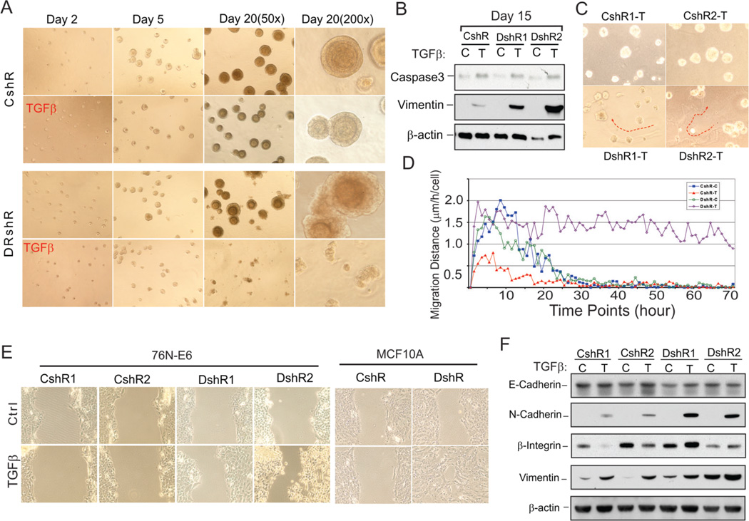
Figure 2. DEAR1 is a negative regulator of TGFβ-induced migration and EMT.
(A) Phase contrast images of DEAR1-shRNA clone (DshR) and control-shRNA clone (CshR) with or without TGFβ (2 ng/ml) in 3D culture at indicated time. Experiments were repeated 2 times, and similar data were obtained. (B) Western analysis of DEAR1-KD clones (DshR) and control clones (CshR) in 3D culture. Cells were collected at the time points indicated with recover buffer (BD) and lysed in 1x SDS sample buffer. (C) Phase contrast images of DEAR1-shRNA clones (DshR1-T and DshR2-T) and control-shRNA clones (CshR1-T and CshR2-T) with or without (data not shown) TGFβ (2 ng/ml) in 3D culture for 5 days. Red arrows trace movement of TGFβ-treated DEAR1 shRNA clones through the matrix. (D) Cell migration distance at each time point comparing 76N-E6 DEAR1-KD clones with or without TGFβ treatment (DshR-T versus DshR-C) and control clones with or without TGFβ treatment (CshR-T versus CshR-C). The values showed are means of 150–200 cells. (E) Wound healing assay of DEAR1-KD (DshR1 and DshR2) and control clones (CshR1 and CshR2) of 76N-E6 (left panel) and MCF10A cells (DshR10A versus control shRNA (CshR10A), right panel) with or without TGFβ treatment (2ng/ml) for 24 hours. (F) Western analysis of EMT markers in 76N-E6 DEAR1-KD (DshR1 and DshR2) and control clones (CshR1 and CshR2) with or without TGFβ treatment (4 ng/ml) for 4 days.

