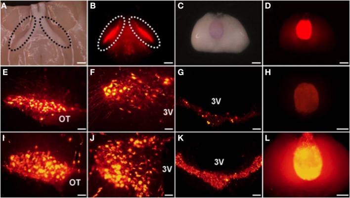
Figure 1.
The mRFP1 fluorescence was clearly observed in ventral parts of the supraoptic nucleus (SON) (A,B) and in the PP (C,D) without cutting. Endogenous florescence of mRFP1 in the SON (E), the paraventricular nucleus (PVN) (F), the median eminence (ME) (G), and the posterior pituitary (PP) (H). Effects of salt loading for 5 days on the mRFP1 fluorescence of the SON (I), the PVN (J), the ME (K), and the PP (L). Under light (A,C) and fluorescent (B,D–L). Scale bars, 1 mm (A–D,H,L) and 0.1 mm (E–G,I–K). OT, Optic tract; 3V, third ventricle. Modified with permission from Figure 1 in Katoh et al. (2011).

