Figure 4.
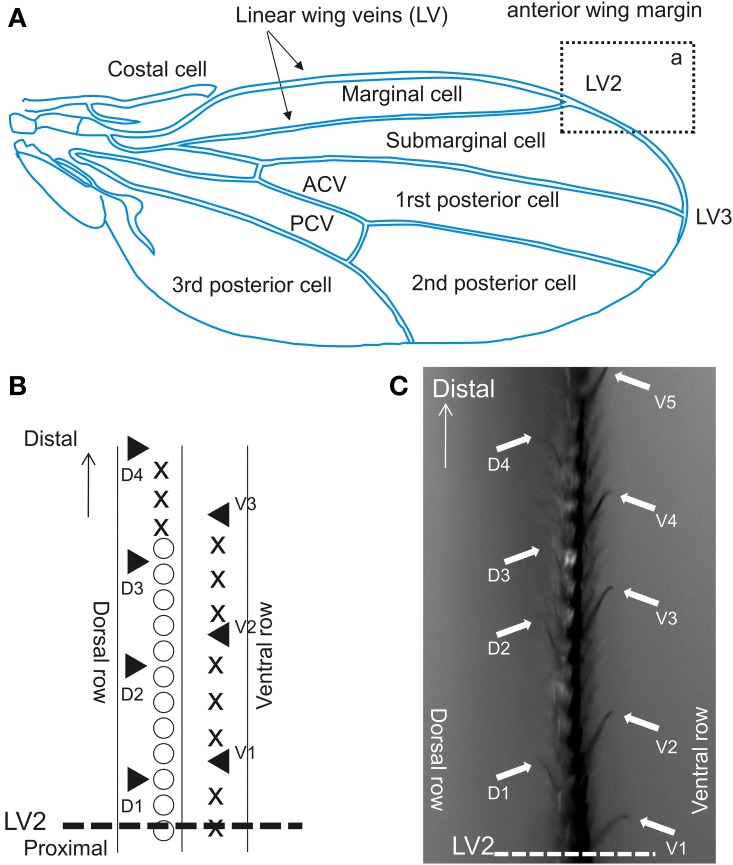
Contact chemoreceptors on the wing. (A) Drosophila wing. ACV, anterior cross vein; PCV, posterior cross vein. The section from which recordings were performed is outlined by a rectangle (a), which displayed at a higher magnification in Figure 2B. (B) Anterior wing margin, with 3 rows of bristles. Symbols on the picture show: ◯, singly innervated stout bristle; X, singly innervated slender bristle; ▲, multiple innervated curved bristles (from which recordings were obtained). (C) Sensilla on the vein area between LV2 and LV3 in rectangle a. Arrows indicate sensilla recorded in this study (▲ in B).

